
Comment est la cholécystectomie laparoscopique
Dec 08, 2021
La cholécystectomie laparoscopique est devenue une technique chirurgicale mature, acceptée par la majorité des patients avec les caractéristiques de moins de traumatisme, moins de douleur et une récupération rapide.
(1) Indications
① Symptomatic gallstones.
② Symptomatic chronic cholecystitis.
③ Gallstone with diameter >3 cm.
④ Filled gallstones.
⑤ Symptomatic and surgically indicated protuberant lesions of the gallbladder.
⑥ The symptoms of acute cholecystitis were relieved after treatment, and there were surgical indications.
⑦ It is estimated that the patient is well tolerated.
(2) Contre-indications relatives
① Acute attack of calculous cholecystitis.
② Chronic atrophic calculous cholecystitis.
③ Secondary choledocholithiasis.
④ History of upper abdominal surgery.
⑤ Fat body.
⑥ External abdominal hernia.
(3) Contre-indication absolue
① Acute cholecystitis with serious complications, such as gallbladder empyema, gangrene, perforation, etc.
② Gallstone acute pancreatitis.
③ With acute cholangitis.
④ Primary common bile duct stones and intrahepatic bile duct stones.
⑤ Obstructive jaundice.
⑥ Gallbladder cancer.
⑦ Protuberant lesions of the gallbladder are suspected to be cancerous.
⑧ Cirrhosis and portal hypertension.
⑨ Middle and late pregnancy.
⑩ Abdominal infection, peritonitis.
Chronic atrophic cholecystitis, gallbladder less than 4.5cm × 1.5cm, wall thickness >0.5 cm (mesure par ultrasons).
Accompagné de maladies hémorragiques et de dysfonctionnement de la coagulation.
Ceux qui ont une fonction incomplète d'organes importants, une opération et une anesthésie difficiles à tolérer, et ceux qui ont un stimulateur cardiaque (l'électrocoagulation et l'électrocoagulation sont interdites).
L'état général est mauvais, il n'est pas adapté à l'opération ou le patient est âgé, il n'y a pas d'indication forte de cholécystectomie, hernie diaphragmatique.
Le champ des indications de la chirurgie laparoscopique s'élargit avec le développement de la technologie. Certaines maladies qui étaient à l'origine des contre-indications à la chirurgie ont également été tentées d'être complétées par la laparoscopie. Si la cholédocholithiase secondaire a été partiellement résolue par chirurgie laparoscopique. Après avoir acquis l'expérience nécessaire, davantage de maladies peuvent être traitées par chirurgie laparoscopique.
(4) Procédure chirurgicale
① Create pneumoperitoneum. Make an arc incision along the lower edge of the umbilical fossa, about 10mm long. If the lower abdomen has been operated on, cut the skin on the upper edge of the umbilical fossa to avoid the original surgical scar.
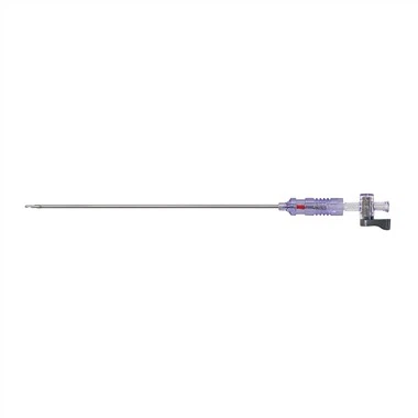
L'opérateur et le premier assistant tiennent chacun une pince à serviette en tissu pour soulever la paroi abdominale des deux côtés de la fosse ombilicale. L'opérateur a tenu l'aiguille de pneumopéritoine (aiguille de Veress) avec le pouce et l'index de sa main droite, a exercé une force sur son poignet et a poignardé la cavité abdominale verticalement ou légèrement obliquement dans la cavité pelvienne.
Au cours du processus de ponction, lorsque l'aiguille traverse le fascia et le péritoine, il y a un sentiment de percée deux fois; Jugez si la pointe de l'aiguille est entrée dans la cavité abdominale. Une seringue avec une solution saline normale peut être connectée. Lorsque la pointe de l'aiguille est dans la cavité abdominale, elle montre une pression négative. Connectez la machine pneumopéritoine. Si la pression de gonflage ne dépasse pas 1,73 kpa, cela indique que l'aiguille de pneumopéritoine est dans la cavité abdominale. Ne gonflez pas trop vite au début. Utilisez un gonflage à faible débit, 1 2L par minute.
At the same time, observe the intraperitoneal pressure on the pneumoperitoneum machine. The pressure during inflation should not exceed 1.73kpa. If it is too high, it indicates that the position of the pneumoperitoneum needle is incorrect, the anesthesia is too shallow and the muscle is not loose enough. Appropriate adjustment should be made. When the abdomen begins to bulge and the liver dullness boundary disappears, it can be changed to high flow automatic inflation until the predetermined value (1.73 2.00kpa) is reached. At this time, the inflation is 3 4L, the patient's abdomen is completely bulged, and the operation can be started.
Lift the abdominal wall with towel pliers at the umbilical pneumoperitoneum needle and puncture with 10mm trocar. The first puncture has a certain "blindness", which is a more dangerous step in laparoscopy. Be extra careful. Rotate the trocar slowly and enter the needle evenly. When entering the abdominal cavity, there is a feeling that the resistance disappears suddenly. Open the closed air valve and gas escapes. This is the success of puncture. Connect the pneumoperitoneum machine to maintain constant pressure in the abdominal cavity. Then put the laparoscope in and puncture at each point under the monitoring of the laparoscope.
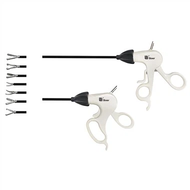
Généralement, percez 2 cm sous le processus xiphoïde et placez un boîtier de 10 mm pour le crochet de décharge, l'applicateur de pince et d'autres instruments ; Ponctionner 2 cm sous le bord costal de la ligne claviculaire moyenne droite ou 2 cm sous le bord externe du droit de l'abdomen et le bord costal du front axillaire avec un trocart de 5 mm respectivement pour mettre en place l'irrigateur et la pince à préhension fixe vésiculaire. À l'heure actuelle, le pneumopéritoine artificiel et les préparatifs sont terminés.
En raison de la fabrication du pneumopéritoine et de la première ponction du trocart, les gros vaisseaux sanguins et les intestins de la cavité abdominale peuvent être accidentellement blessés et il n'est pas facile de les trouver pendant l'opération. Récemment, de nombreuses personnes ont fait une petite ouverture dans l'ombilic pour trouver le péritoine et mettre directement le trocart dans la cavité abdominale pour le gonflage. Après la fabrication réussie du pneumopéritoine, l'opération a commencé.
② Dissect the Calot triangle. Grasp the neck of gallbladder or Hartmann's bursa with grasping forceps and traction to the upper right. It is best to draw the cystic duct perpendicular to the common bile duct in order to clearly distinguish the two, but pay attention not to draw the common bile duct into an angle. The serous membrane on the cystic duct was cut with an electrocoagulation hook, the cystic duct and cystic artery were passively separated, and the common bile duct and common hepatic duct were distinguished. Since it is close to the common bile duct, electrocoagulation should be used as little as possible to avoid accidental injury to the common bile duct. Use the electrocoagulation hook to separate the cystic duct upstream and downstream, and see the relationship between the cystic duct and the common bile duct. Place the titanium clip as close to the gallbladder neck as possible. There should be sufficient distance between the two titanium clips. The titanium clip should be at least 0.5cm away from the common bile duct. Cut between the two titanium clips with scissors, and do not use electric cutting or electrocoagulation to prevent damage to the common bile duct due to heat conduction. Then find the cystic artery behind it and cut it with titanium clip. After cutting off the gallbladder artery, do not pull hard to avoid breaking the gallbladder artery, and pay attention to the posterior branch of the gallbladder. Carefully peel off the gallbladder, electrocoagulation or hemostasis with titanium clip.
③ Cholecystectomy. Clamp the gallbladder neck and pull it upward, carefully peel it off along the gallbladder wall, and the assistant should assist in pulling to make the gallbladder and liver bed have a certain tension. Completely peel off the gallbladder and place it on the upper right side of the liver. The liver bed was hemostatic by electrocoagulation, carefully rinsed with normal saline, and checked for bleeding and bile leakage (a piece of gauze was disposed at the hepatic hilum, and checked for bile staining after removal). After absorbing all the water in the abdominal cavity, transfer the laparoscope to the lower sleeve of the xiphoid process and give way to the umbilical incision, so that the gallbladder containing stones greater than 1cm can be taken out from the umbilical incision with loose structure and easy expansion. If the stones are small, they can also be taken out from the puncture hole under the xiphoid process.
④ Remove the gallbladder. Put the toothed claw forceps into the abdominal cavity from the cannula at the umbilicus, grasp the residual end of the cystic duct under monitoring, slowly drag the gallbladder into the cannula sheath and pull it out together with the cannula sheath. When grasping the gallbladder, pay attention to placing the gallbladder on the liver to avoid accidental injury to the intestinal canal by sharp forceps. If the stone is large or the tension of the gallbladder is high, do not pull it out with force to avoid rupture of the gallbladder and leakage of stones and bile into the abdominal cavity. At this time, the incision can be enlarged with vascular forceps and taken out, or the incision can be expanded to 2.0cm with an expander. If the stone is too large, the incision can be extended. If bile leaks into the abdominal cavity, wet gauze shall be used to enter from the umbilical incision to suck up the bile.
Si la pierre est trop grosse pour être retirée de l'incision, vous pouvez également ouvrir d'abord la vésicule biliaire, aspirer la bile dans la vésicule biliaire avec un aspirateur et la retirer une par une après avoir écrasé la pierre avec une pince. Si une pierre tombe dans la cavité abdominale, retirez-la. Après avoir vérifié qu'il n'y a pas de sang et de liquide dans la cavité abdominale, retirez le laparoscope, ouvrez la valve de la canule pour évacuer le gaz carbonique dans la cavité abdominale, puis retirez la canule. L'incision avec une canule de 10 mm est suturée avec un fil fin comme couche de fascia pour 1 à 2 points de suture, et chaque incision est fermée avec un film adhésif stérile.
(5) Complications majeures
① Bile duct injury. Bile duct injury is one of the most common and serious complications of laparoscopic cholecystectomy.
L'incidence des lésions des voies biliaires et des fuites biliaires est d'environ 10 %. Il faut y prêter suffisamment d'attention. Elle est principalement due à l'anatomie peu claire du triangle de Calot, en particulier au manque de vigilance vis-à-vis de la variation commune du cholédoque ou du canal cystique. Lors de la séparation du canal cystique, le canal biliaire a été thermiquement endommagé par inadvertance, il n'y a pas eu de fuite de bile pendant l'opération, et la nécrose et la chute du tissu dans la zone endommagée thermiquement après l'opération pourraient également provoquer une fuite de bile. De plus, il y a souvent de gros canaux biliaires vagaux dans le lit de la vésicule biliaire. L'électrocoagulation peropératoire ne peut pas coaguler complètement et une fuite de bile peut également se former. Les principales manifestations des lésions des voies biliaires sont des douleurs abdominales hautes sévères, une forte fièvre et une jaunisse. Les patients présentant des manifestations typiques sont généralement traités à temps après l'opération ; Cependant, quelques patients n'ont présenté qu'une distension abdominale, un manque d'appétit, une faible fièvre et une exacerbation progressive. Ces patients doivent être étroitement surveillés. Il a été rapporté que l'accumulation de bile intra-abdominale a été trouvée quelques mois après l'opération. Juger s'il y a une fuite biliaire dépend principalement de l'échographie ou de la TDM, puis confirmé par une ponction à l'aiguille fine sous la direction de l'échographie ou de la TDM ou de l'hépatocholangiographie radionucléide.
② Vascular injury. One is massive hemorrhage caused by needle tip injury to abdominal aorta, iliac artery or mesenteric vessels during pneumoperitoneum and trocar placement. There are many reports of death caused by trocar puncture. Therefore, after successful pneumoperitoneum, laparoscopy should peep the whole abdomen once to prevent missing vascular injury.
L'autre est l'anatomie peu claire du portail hépatique ou le mauvais serrage de l'artère hépatique droite ou de l'artère hépatique appropriée en raison d'un saignement de l'artère de la vésicule biliaire. Il existe également des rapports de lésions de la veine porte au cours de l'anatomie. Des cas de nécrose hépatique droite causée par un mauvais clampage de l'artère hépatique ont été signalés.
③ Intestinal injury. Intestinal injuries are mostly accidental injuries caused by electrocoagulation, mainly because the electrocoagulation hook is not placed in the TV monitoring picture and is not found. Abdominal pain, abdominal distention and fever occur after operation, resulting in serious peritonitis, and its mortality is high.
④ Postoperative intraperitoneal hemorrhage. Postoperative intraperitoneal hemorrhage is also one of the serious complications of laparoscopic surgery. The injured parts are mainly the blood vessels near the gallbladder, such as hepatic artery, portal vein and abdominal aorta or vena cava during periumbilical puncture. The manifestations were hemorrhagic shock, abdominal bulge and peripheral circulatory failure. Open surgery should be performed immediately to stop bleeding.
⑤ Subcutaneous emphysema. The causes of subcutaneous emphysema are as follows: first, when making pneumoperitoneum, the pneumoperitoneum needle did not penetrate the abdominal wall, and high-pressure carbon dioxide entered the subcutaneous; Second, due to the small skin incision, the trocar is embedded very tightly, and the puncture hole of the peritoneum is relatively loose. During the operation, carbon dioxide gas leaks into the lower skin layer of the abdominal wall. Postoperative examination can find abdominal subcutaneous twisting pronunciation, generally without special treatment.
⑥ Others. Such as incisional hernia, incisional infection and abdominal abscess.